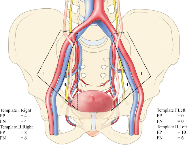
Fig. 2.
Schematic overview of the false-positive and false-negative lymph-node findings when comparing 18F-DCFPyL PET/CT with histopathology, classified according to the templates used in the extended pelvic lymph-node dissection. Template I (green) involves lymph nodes surrounding the arteria iliaca externa. Template II (red) involves lymph nodes surrounding the arteria iliaca interna, and the nervus obturatorius. FP, false-positive lymph-node finding; FN, false-negative lymph-node finding

