Figure 1.

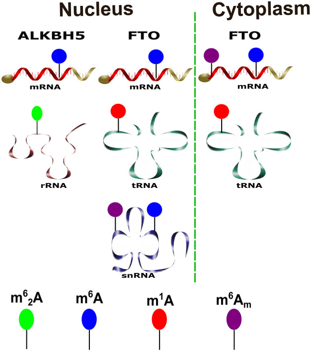
Various physiological substrates of the m6A demethylases. A schematic diagram shows substrates of ALKBH5 and FTO and their distribution in both cytoplasm and nucleus to the different forms of RNAs inside the cell.

Various physiological substrates of the m6A demethylases. A schematic diagram shows substrates of ALKBH5 and FTO and their distribution in both cytoplasm and nucleus to the different forms of RNAs inside the cell.