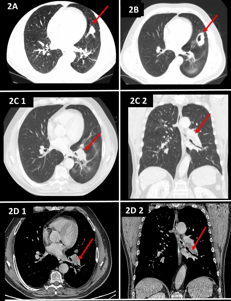
Fig. 2.
a High-resolution CT image revealing a large nodule in the lower lobes. b High-resolution CT image revealing a cavitated nodule. c CT pulmonary window revealing enlarged lymph nodes in the hilus of the left lung that impress the bronchi to the lower lobe, which, combined with the presence of subpleural nodules, aroused suspicion of a proliferative process (2C1 axial scan, 2C2 coronal scan). d CT soft tissue window revealing a pathological mass in the hilus of the left lung (2D1 axial scan, 2D2 coronal scan)

