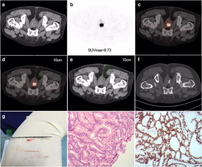
Fig. 4.
PSMA-TB was performed in a 73-year-old man (without rectal access) with serum PSA 35.6 μg/ml who had undergone rectal surgery for rectal carcinoma before enrolment in the study. a Normal CT; b PSMA-avid lesion (SUVmax 8.73); and c fusion image. Puncture path and angle pre-simulated on d fusion image and e CT image in the same scanning plane with a puncture depth of 10.0 cm. f CT image of transgluteal biopsy needle guided into the target lesion. g Three specimens were taken using the co-axial needle technique with lengths of 0.8, 1.0 and 2.0 cm. h Specimen was positive for PCa (haematoxylin and eosin staining, 10 × 10) with Gleason score 7 (4 + 3). i Positive PSMA expression was confirmed by EnVision immunostaining

