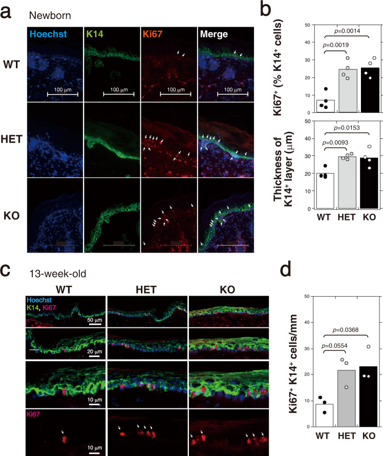
Fig. 2. Proliferation of keratinocytes is accelerated by downregulation of CS-C.
a Immunofluorescence labeling of skin from newborn C6st-1 WT, C6st-1 HE, and C6st-1 KO mice was performed using anti-K14 and anti-Ki67 antibodies. Nuclei are counterstained with Hoechst33342. b Quantification of Ki67-positive cells in K14-positive basal layers, and measurement of epidermal thickness in K14-positive basal layer of C6st-1 WT (n = 4), C6st-1 HE (n = 4), and C6st-1 KO mice (n = 4). Multiple random vertical lines perpendicular to the epidermal border were measured. From the mean thickness of these lines, epidermal thickness in K14-positive basal layer was calculated. c Immunofluorescence labeling of tail skin from 13-week-old C6st-1 WT, C6st-1 HE, and C6st-1 KO mice was performed using anti-K14 and anti-Ki67 antibodies. Nuclei are counterstained with Hoechst33342. d Numbers of Ki67-positive cells within 1 mm of K14-positive basal layers in C6st-1 WT (n = 3), C6st-1 HE (n = 3), and C6st-1 KO mice (n = 3). The number of Ki67-positive cells per mm of basal layer length was counted manually using a digital imaging software (Photoshop CS6). Statistical significance was determined using one-way ANOVA with Tukey’s HSD test.

