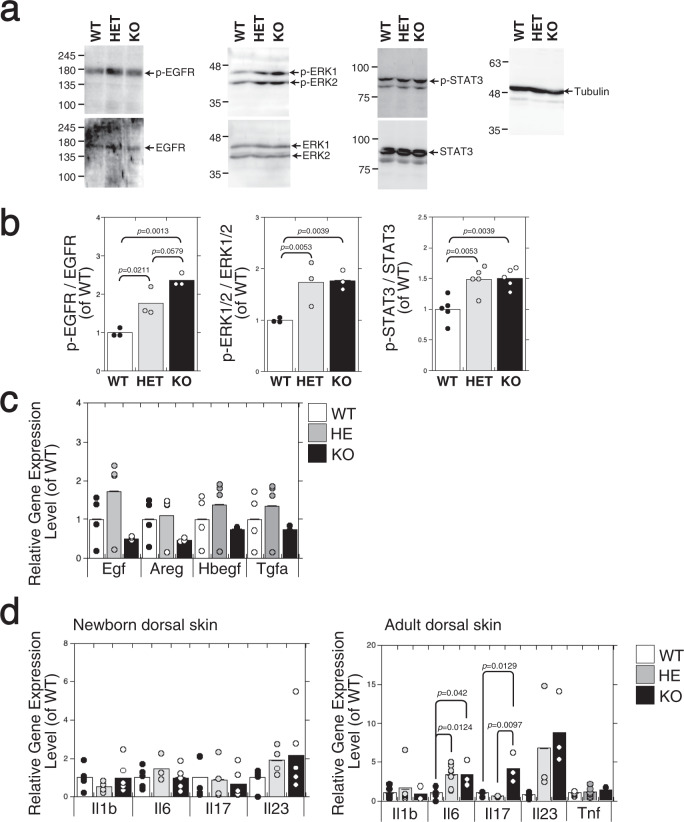
Fig. 4. The expression of phospho-EGFR, phospho-ERK1/2, and phospho-STAT3 is augmented in the epidermis of C6st-1 HE and C6st-1 KO mice compared with that in C6st-1 WT mice.
a Proteins extracted from the epidermis of newborn C6st-1 WT, C6st-1 HE, and C6st-1 KO mice were analyzed by immunoblotting using anti-phospho-EGFR (p-EGFR), anti-EGFR, anti-phospho-ERK1/2 (p-ERK1/2), anti-ERK1/2, and anti-phospho-STAT3 (p-STAT3), anti-STAT3, and anti-tubulin antibodies. b Densitometric analysis of the levels of phospho-EGFR, phospho-ERK1/2, and phospho-STAT3 compared with levels of total EGFR, ERK1/2, and STAT3 is shown. Bars represent the means ± S.D. from three independent biological replicates. Statistical significance was determined using one-way ANOVA with Tukey’s HSD test. c The expression levels of EGFR ligands (Egf, Areg, Hbegf, and Tgfa) in C6st-1 WT, C6st-1 HE, and C6st-1 KO keratinocytes were analyzed using real-time PCR (n = 4). Values are mean ± S.D. d Expression levels of pro-inflammatory cytokines (Il1b, Il6, Il17, and Il23) in dorsal skin of C6st-1 WT, C6st-1 HE, and C6st-1 KO newborn and adult (13-week-old) mice were analyzed using real-time PCR (n = 3–7). Values represent the mean ± S.D. Statistical significance was determined using one-way ANOVA with Tukey’s HSD test.

