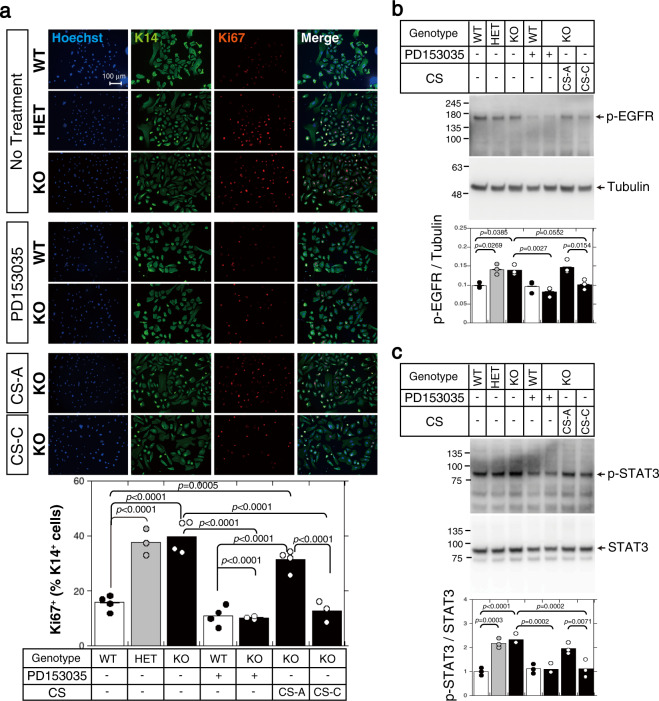
Fig. 5. EGFR-dependent keratinocyte proliferation is regulated by CS-C.
a Keratinocytes (1 × 10⁵ cells/well), isolated from C6st-1 WT, C6st-1 HE, and C6st-1 KO epidermis, were cultured in the presence or absence of 5 μM PD153035, 100 μg/ml CS-A, and 100 μg/ml CS-C. Cells were immunolabeled using anti-K14 and anti-Ki67 antibodies. The graph below indicates quantification of Ki67- and K14-double positive cells. Values are mean ± S.D. with n = 3-4 per group. Statistical significance was determined using one-way ANOVA with Tukey’s HSD test. Keratinocytes were treated with 5 μM PD153035, 100 μg/ml CS-A, and 100 μg/ml CS-C as indicated. Cells were lysed and subjected to immunoblotting using b anti-phospho-EGFR and anti-tubulin antibodies, and c anti-phospho-STAT3 and anti-STAT3 antibodies. Tubulin and STAT3 were used as loading controls. The graphs below the blots show relative expression levels of phospho-EGFR and phosphor STAT3 in C6st-1 WT, C6st-1 HE, and C6st-1 KO keratinocytes subjected to the indicated treatments. Values are mean ± S.D. with n = 3 per group. Statistical significance was determined using one-way ANOVA with Tukey’s HSD test.

