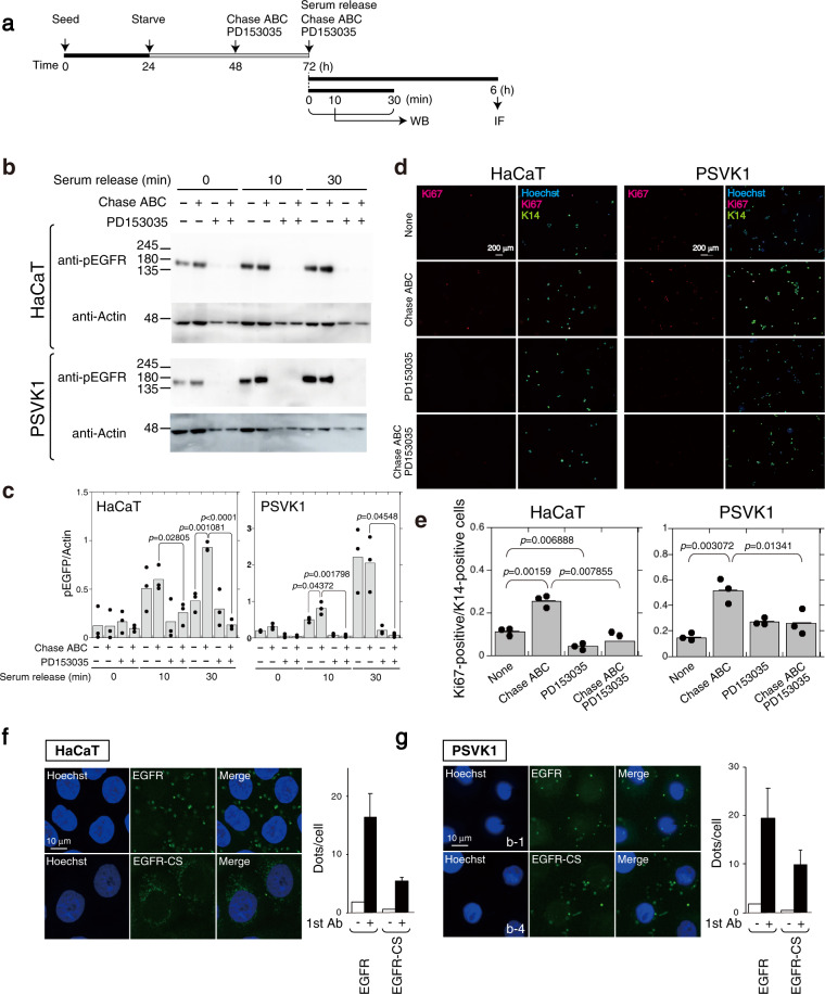
Fig. 7. Proliferation of cells from human keratinocyte clone lines, HaCaT and PSVK1, is regulated by CS.
a Schematic diagram of experimental workflow. b HaCaT cells (2 × 10⁴ cells/dish) and PSVK1 cells (1.7 × 10⁵ cells/dish) were seeded, and starved for 48 h prior to allowing cell cycle progression via addition of serum in the presence of 2.5 munits/mL of Chase ABC and 5 μM PD153035, and culturing cycling cells for the indicated time. Proteins extracted from HaCaT and PSVK1 cells were analyzed by immunoblotting using anti-phospho-EGFR (p-EGFR), and anti-EGFR antibody. c Densitometric analysis of the levels of phospho-EGFR compared with levels of actin is shown. Bars represent the means ± S.D. from three independent biological replicates. Statistical significance of differences was determined using Student’s t test. d HaCaT (1 × 10⁴ cells/well) and PSVK1 cells (2.5 × 10⁴ cells/well) were cultured in the presence or absence of Chase ABC (2.5 munits) and PD153035 (5 μM), and stained with anti-K14 and anti-Ki67 antibody. Nuclei were counterstained with Hoechst33342. e Quantification of Ki67- and K14-double positive keratinocytes is shown. Values are mean ± S.D. from three independent biological replicates. Statistical significance was determined using Student’s t test. Direct interactions between EGFR and CS-C (EGFR-CS) and EGFR homodimers in HaCaT (f) and PSVK1 (g) cells were detected by PLA assay. The graph shows means ± S.D. of EGFR homodimer and EGFR-CS signals (green dots)/cell counted from three independent experiments.

