Figure 3.
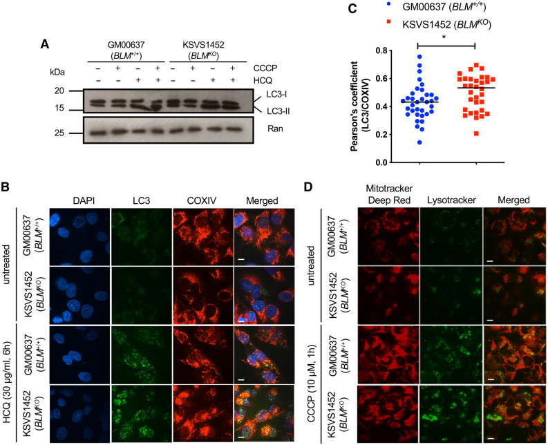
BLM-deficient cells are proficient in autophagy. (A) Western blot analysis of GM00637 (BLM+/+) and KSVS1452 (BLMKO) cells for levels of cytosolic LC3-I and autophagosomal marker LC3-II. Both, cytosolic LC3-I and membrane-bound LC3-II, were detected with LC3 antibody G-9 (SCBT). Cells were treated with 10 µM CCCP for 6 h to induce autophagy and/or with 30 µg/ml HQC for 6 h to interrupt autophagic flux. (B) Confocal microscopy images of fixed GM00637 (BLM+/+) and KSVS1452 (BLMKO) cells immunostained for LC3 (MAP LC3β-G-9 (SCBT) and COXIV (Cell Signaling), and stained with DAPI. Cells were treated with 30 µg/ml HCQ for 6 h to interrupt autophagic flux. Scale bars 10 µm. (C) Quantification of colocalization of antibody staining for LC3 and COXIV in HCQ-treated cells imaged as in panel B. 30 cells from BLM-proficient GM00637 cells and BLM-deficient KSVS1452 cells were analyzed. Significance of differences was determined by a Student’s t test and is reported as *p ≤ 0.05. (D) Confocal microscopy images of BLM-proficient GM00637 cells and isogenic BLM-knockout cells KSVS1452 stained with MitoTracker Deep Red and Lysotracker in the presence and absence of CCCP. Scale bars 10 µm.

