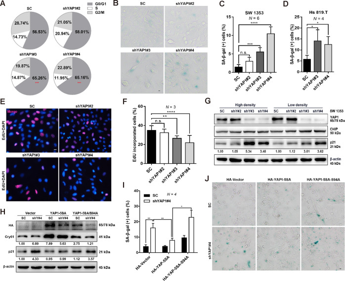
Fig. 2. YAP1/TEAD controls cell cycle exit and induces cellular senescence.
A Cell cycle distribution analysis in the groups of control shRNA and YAP1-specific shRNAs (shYAP#2, #3, #4). B, C Control shRNA or YAP1-depleted SW 1353 cells were stained with SA-β-gal, and the representative images are shown in (B); the percentage of SA-β-gal-positive cells was quantified and shown in (C), scale bars: 10 μm. D Hs 819.T cells were infected with lentivirus of control shRNA or YAP1-specific shRNAs (shYAP#3, #4), and then the percentage of SA-β-gal-positive cells was quantified. E, F YAP1 depletion inhibits cell proliferation. Cell proliferation was determined by the EdU incorporation assay (E), scale bar: 10 μm. DAPI was used as a nuclear counterstain. The percentage of cells that incorporated EdU was quantified (F). G Knockdown of YAP1 induces the increase of p21 expression, independent of cell density. Expression of the indicated proteins was determined by IB. H–J YAP1-TEAD plays a role in regulating CHS cell senescence. control shRNA or YAP1-depleted SW 1353 cells were transiently transfected with HA-vector (Vector), HA-YAP1-5SA (YAP1-5SA), or HA-YAP1-5SA/S94A (YAP1-5SA/S94A) for 72 h, and then the protein levels of HA, Cyr61, and p21 were determined by IB (H). The percentage of SA-β-gal-positive cells was quantified (I), and representative images are shown in (J); scale bars: 10 μm. Data are presented as the mean ± SD of at least three independent experiments in (A, C, D, F, I). One-way ANOVA followed by Dunnett’s test was applied for (A, C, D, F). Two-way ANOVA followed by Tukey’s test for (I). n.s. nonsignificant, *P < 0.05, **P < 0.01, ***P < 0.001, ****P < 0.0001.

