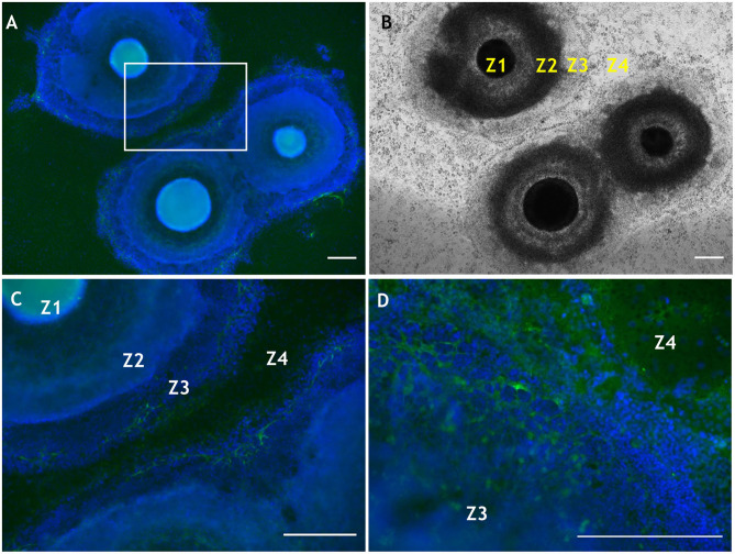
Figure 4.
Localization of CS using antibody 7D4 within developing SEAM cultures at weeks 4 and 6 of differentiation. (A) Wide-field view of three SEAM colonies at week 4 of differentiation, two of which are beginning to merge (7D4 is shown in green and nuclear Hoechst-334 staining in blue). (B) Bright-field image of a SEAM at week 4 of differentiation with each zone (Z) indicated. (C) Higher magnification view of CS immunolocalisation in a selected region in (A). (D) High magnification of a SEAM at week 6 of differentiation indicating a widespread deposition of CS. Scale bars = 100 μm. Methods and negative control images with primary antibodies omitted are provided as Supplementary Material.

