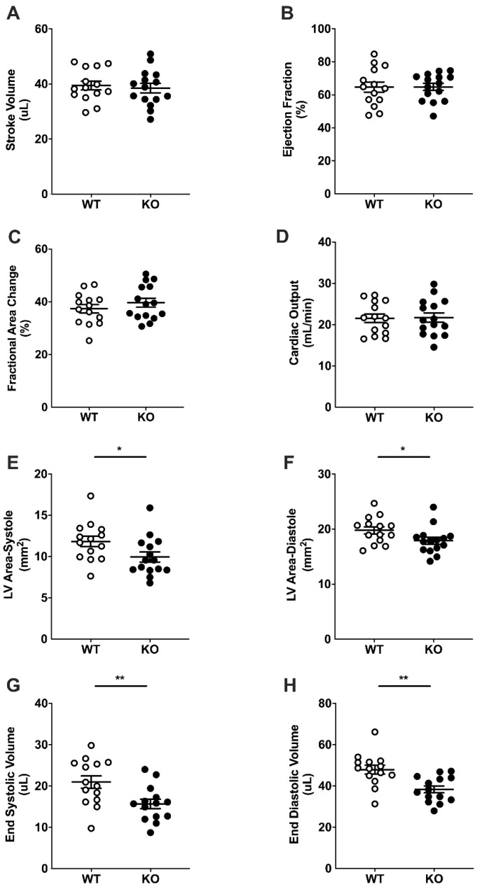
Figure 3.
Basal cardiac dimensions and function by ultrasound echocardiography. Stroke volume (SV, A), ejection fraction (EF, B), fractional area change (FAC, C), cardiac output (CO, D), left ventricular systolic (E) and diastolic (F) areas, end systolic volume (ESV, G) and end diastolic volume (EDV, H) in 8-week-old female WT (white, n = 14) and KO (black, n = 14) mice. Student’s t-test. Data are mean ± SEM. * p < 0.05, ** p < 0.01.

