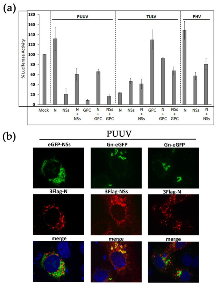
Figure 3.
Effect of co-expressing different hantaviral proteins on RIG-I-induced IFN promoter activity and subcellular localization in transfected cells. (a) HEK293T cells were transfected with 300 ng of plasmid encoding either 3×Flag-N, 3×Flag-NSs, or StrepTag-GPC individually or in combination (N+NSs, N+GPC or NSs+GPC) using 150 ng of each plasmid. Inhbitory effect of the constructs was measured on RIG-I-induced IFNβ-Luc activity, as described in Figure 1. In (b) such combinations of PUUV constructs were co-transfected in VeroE6 cells. Intracellular localization of N with eGFP-NSs (left panels), and of Gn-eGFP with NSs (middle panels) or with N (right panels) were visualized by immunofluorescence. Proteins directly coupled to eGFP appeared in green, and N or NSs tagged proteins stained with anti-Flag mAb, then anti IgG-Alexa 555, appeared in red. The nuclei were labeled in blue with DAPI. The merge panels are shown at the bottom of the figures and co-localizing proteins appearing in yellow. The images obtained using an objective x63 are at the same magnification.

