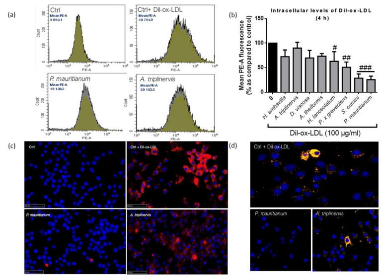
Figure 3.
Dil-ox-LDL uptake by RAW 264.7 macrophages in the presence of medicinal plant decoctions analyzed by flow cytometry (a,b), nanozoomer scanner (c) and confocal microscopy (d), data shown in (b) are means ± SD of three independent experiments. # p < 0.05, ## p < 0.01, ### p < 0.005 as compared to cells treated with Dil-ox-LDL (0 in black). Blue color refers to DAPI (4′,6-diamidino-2-phenylindol) nuclear staining and red/yellow colors refer to Dil-ox-LDL, respectively, observed by fluorescence and confocal microscopy.

