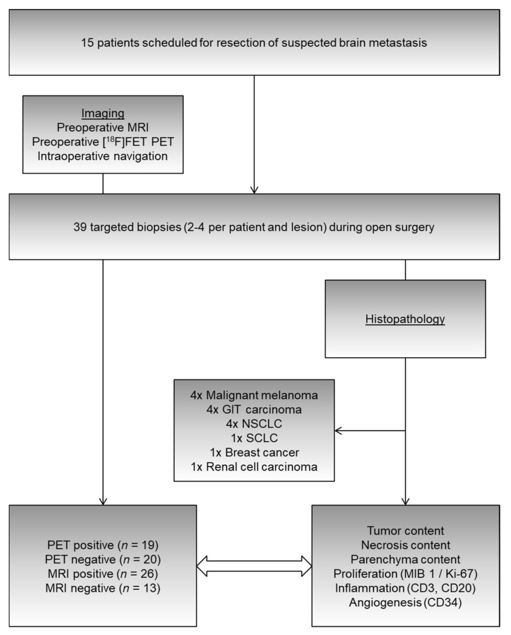
Figure 2.
From 15 patients undergoing resection of suspected brain metastases, 39 targeted (i.e., navigated) biopsies were obtained from parts of the tumor that were MRI-positive, MRI-negative, PET-positive, or PET-negative (cf. Figure 1). Histopathological evaluation permitted the investigation of relationships between imaging and tissue properties, such as tumor/necrosis/parenchyma content (percentages), proliferation indices (MIB 1/Ki-67), and markers of inflammation (CD3 = T-lymphocyte infiltration, CD20 = B-lymphocyte infiltration) as well as angiogenesis (CD34). GIT: gastrointestinal tract; NSCLC: non-small cell lung cancer; SCLC: small-cell lung cancer; CD: cluster of differentiation.

