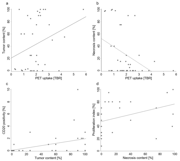
Figure 3.
Relationships of PET imaging and histological tumor properties. In brain metastases, 18F-FET uptake imaged by PET (measured as mean tumor-to-background ratio (TBRmean) of the target volume) increased with tumor content (a; rs = 0.3, p = 0.045) and decreased with necrosis content (b; rs = −0.4, p = 0.014). B-lymphocyte infiltration (measured by CD20 positivity) increased with tumor content (c; rs = 0.5, p = 0.002) and necrosis content increased with proliferation (d; rs = 0.5, p = 0.002). Note that while scatterplots and linear regression lines are shown to illustrate the data, correlations were analyzed using Spearman’s rank correlation coefficient rs, as tumor content, necrosis content, and CD20 positivity were not normally distributed.

