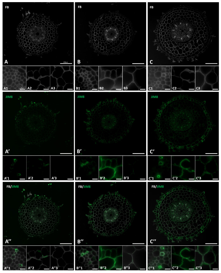
Figure 4.
Immunolocalization of the JIM8 epitope in cross-sections of the “salt stressed” Brachypodium roots. (A,A′,A″) control, (B,B′,B″) 100 mM NaCl, (C,C′,C″) 200 mM NaCl. Fragments of vascular cylinder are marked by the numeral 1, rhizodermis and exodermis by the numeral 2, and cortex cells by the numeral 3. FB—fluorescent brightener. The green colour shows epitope occurrence. Scale bars for A,A′,A″, B,B′,B″, C,C′,C″ represent 100 µm and for all other photomicrographs: 10 µm.

