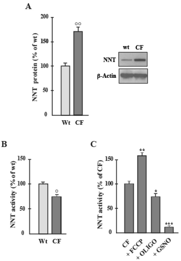
Figure 1.
NNT protein level and activity in airway cells. (A) NNT protein expression in Wt and CF cells was detected by Western blot analyses. The blot incubated for each antibody was stripped and reprobed with anti ß-actin antibody. Protein amount was expressed, after normalization with respect to corresponding β-actin, as a percentage of the content inWt cells to which a value of 100 was CCCP (3 μmol/L), OLIGO (1.5 ng/ml) and GSNO (1 mmol/L) were added 15 min before activity determination. Values were subjected to statistical analysis (Wt, n = 4; CF, n = 5): (B), ° p < 0.05 when comparing CF with the Wt-samples); (C), * p < 0.05, ** p < 0.01, *** p < 0.001 when comparing untreated cells with the cells in the presence of compounds).

