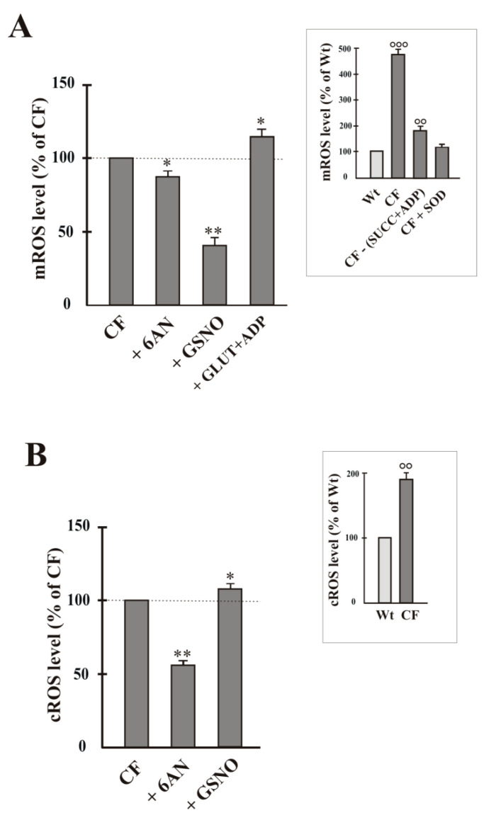
Figure 4.
ROS level in airway cells. Mitochondrial and cytosolic O2−• production was detected by using the MitoSox dye (A) and according to the adrenochrome method (B), respectively (see Method Section). The O2−• level value was expressed as % of control, i.e., sample in the absence of compound, to which value 100 was given. Cells were incubated for 15 min in the absence or presence of 6AN (5 mmol/L), GSNO (1 mmol/L), GLUT (10 mmol/L) + ADP (1 mmol/L). Values were subjected to statistical analysis (for both Wt and CF, n = 4). * p < 0.05; ** p < 0.01 when comparing untreated CF cells with the same cells in the presence of compounds. In the Insets, ROS level value was expressed as % of control (Wt) to which value 100 was given. °° p < 0.01; °°° p < 0.001 versus Wt.

