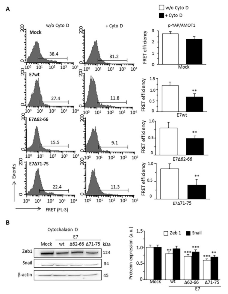
Figure 6.
Effect of Cytochalasin D on p-YAP/AMOT1 interaction and expression of EMT markers. (A) Quantitative evaluation of p-YAP/AMOT1 molecular association by FRET technique, as revealed by flow cytometry analysis restricted to pAmCyan-positive cells treated or not with CytoD (1 mM for 4 h). Numbers indicate the percentage of FL3 positive events obtained in one experiment representative of three. Bar graphs on the right show FRET efficiency calculated according to the Riemann’s algorithm. Data are reported as mean ± SD from three independent experiments. ** p < 0.01 vs. Mock transfected cells. (B) Western blot analysis of the expression of the EMT markers Zeb1 and Snail in C33A expressing E7wt or E7 mutated treated with CytoD. β-actin determination was used as loading controls. Bar graph (right) shows relative densitometry quantitation of each protein normalized to β-actin obtained in three independent experiments and reported as mean ± SD. ** p < 0.01, *** p < 0.001 vs the same sample treated with CytoD. Uncropped western blot images for Figure 6B are available in Supplementary Figure S9.

