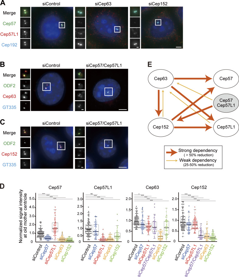
Figure 6.
Interdependency of centrosomal localization of Cep57, Cep57L1, Cep63, and Cep152. (A) The centrosomal localization of Cep57 and Cep57L1 was dependent on Cep63 and Cep152. HeLa cells were treated with siControl, siCep63, or siCep152 and immunostained with antibodies against Cep57 (green), Cep57L1 (red), and Cep192 (cyan). (B) The centrosomal localization of Cep63 was partially dependent on Cep57 and Cep57L1. HeLa cells were treated with siControl or siCep57/Cep57L1 and immunostained with antibodies against ODF2 (green), Cep63 (red), and GT335 (cyan). (C) The centrosomal localization of Cep152 was partially dependent on Cep57 and Cep57L1. HeLa cells were treated with siControl or siCep57/Cep5L1 and immunostained with antibodies against ODF2 (green), Cep152 (red), and GT335 (cyan). (D) Beeswarm plots piled on boxplots represent the normalized signal intensity of Cep57, Cep57L1, Cep63, and Cep152 at the old mother centrioles upon the indicated siRNAs (n = 50). (E) Schematic of the dependency of the centrosomallocalization among Cep57, Cep57L1, Cep63, and Cep152. The wide arrows indicate strong dependency, defined as >50% reduction of the signal intensity, and narrow arrows indicate weak dependency, defined as 25–50% reduction of the signal intensity. All scale bars, 5 µm in the low-magnification view, 1 µm in the inset. The normal distribution of datasets was confirmed using the Kolmogorov–Smirnov test in D. Tukey’s multiple comparisons test was used in D to obtain P value. *, P < 0.05; ***, P < 0.001; NS, P > 0.05.

