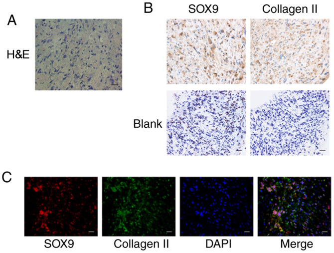
Figure 2.
Chondrogenic differentiation of gb-MSCs in micromass cultured for 21 days. (A) H&E staining of gb-MSCs. (B) SOX-9 and collagen II immunohistochemical staining of cartilage nodules. (C) Double staining for SOX-9 (red) and Collagen II (green) demonstrates that gb-MSCs have the capacity to differentiate into cartilage nodules. Magnification, ×400; scale bar, 25 µm. gb-MSC, glioma-associated mesenchymal stem cells; H&E, hematoxylin and eosin; SOX-9, SRY-Box transcription factor 9.

