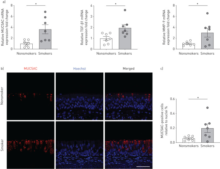
FIGURE 1.
Freshly isolated human bronchial epithelial cells (P0 HBECs) from nonsmokers and smokers. a) Quantitative mRNA expression of MUC5AC, transforming growth factor (TGF)-β1 and matrix metalloproteinase (MMP)-9 of P0 HBECs from nonsmokers and smokers. Data are shown as relative to glyceraldehyde 3-phosphate dehydrogenase (GAPDH) and nonsmokers (n≥6 donors for each group). b) Representative confocal images of immunofluorescent staining of bronchial tissue from a nonsmoker and a smoker. MUC5AC in red; nuclei stained with Hoechst in blue. Scale bar=50 μm. c) Quantification of MUC5AC-positive cells relative to nuclei (n=7 from ≥3 donors for each group). *: p<0.05 using t-test after passing Shapiro–Wilk normality test.

