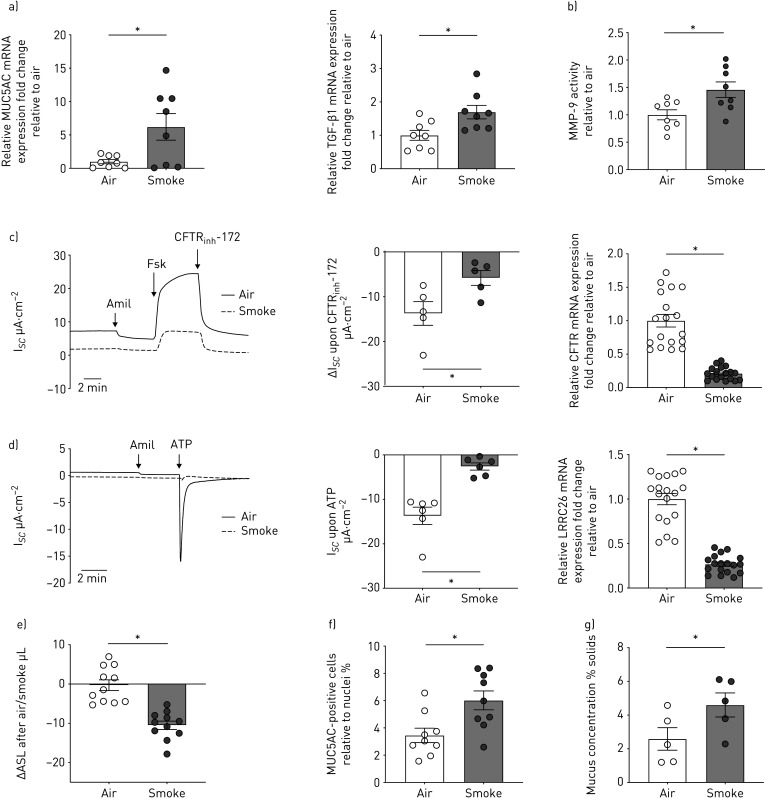
FIGURE 3.
Fully re-differentiated human bronchial epithelial cells (P1 HBECs) from nonsmokers exposed to cigarette smoke (smoke). a) Quantitative mRNA expression of MUC5AC and transforming growth factor (TGF)-β1 in P1 HBECs 24 h after exposure to room air or smoke (24 puffs). Data are shown as relative to glyceraldehyde 3-phosphate dehydrogenase (GAPDH) and air control (n=8 donors for each group). b) Matrix metalloproteinase (MMP)-9 activity assay from PBS washes of P1 HBECs from nonsmokers 24 h after exposure to room air or smoke (24 puffs). Data are shown as relative to air control (n=8 from 4 donors for each group). c) Left panel: representative cystic fibrosis transmembrane conductance regulator (CFTR) trace measured by short circuit current changes upon CFTRinh172 (10 µM) application after 10 µM forskolin stimulation 4 h after exposure to room air or smoke (represented as ΔIsc upon CFTRinh172; thus, decreases indicate enhanced CFTR function) (n=5 donors for each group). Middle panel: quantification of CFTR currents upon CFTRinh172. Right panel: CFTR mRNA expression. Data are shown as relative to GAPDH and air control (n=18 from 6 donors for each group). d) Left panel: representative voltage-dependent potassium (BK) trace and quantification of currents measured upon ATP stimulation 4 h after exposure to room air or smoke (represented as ΔIsc with decreases indicating better BK function). n=6 donors for each group. Right panel: LRRC26 mRNA expression (γ subunit of BK critical for BK function). Data are shown as relative to GAPDH and air control (n=18 from 6 donors for each group). e) Airway surface liquid (ASL) volumes represented as change in volume between 1 and 4 h after air or smoke exposure (n=11 donors for each group). f) Quantification of MUC5AC-positive cells relative to nuclei 24 h after air or smoke exposure (n=9 from 3 donors for each group). g) Mucus concentration depicted as % mucus solids measured 24 h after air or smoke exposure (n=5 donors for each group). *: p<0.05, t-test after passing Shapiro–Wilk normality test for all except for the BK data (Mann–Whitney U-test).

