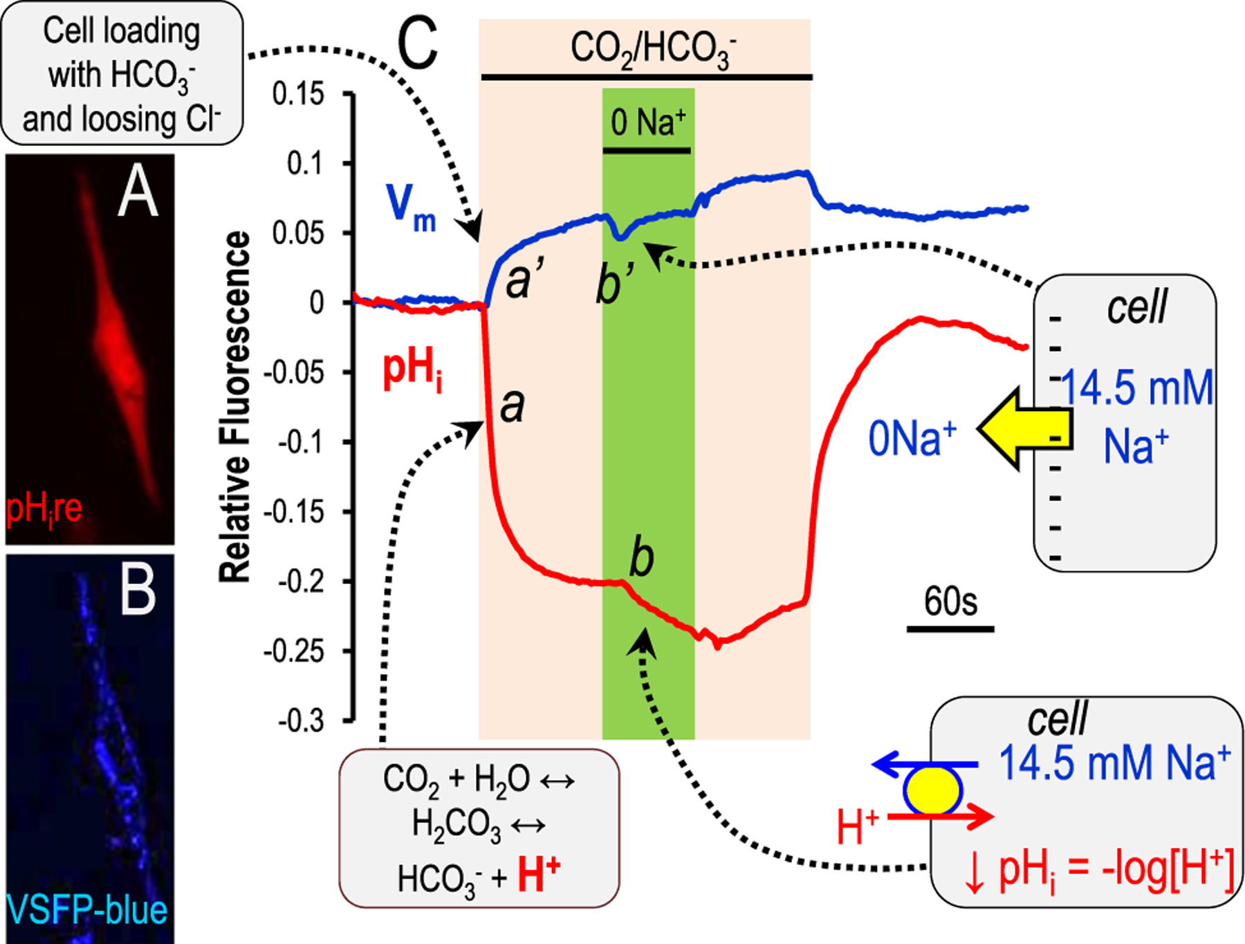
Figure 6.

Genetically encoded pH sensors in mammalian cells. The two trace lines (blue and red) illustrate relative fluorescent responses of TM5 (normal human trabecular meshwork) cells transfected with two genetically encoded sensors. Blue is VSFP blue (lower inset) and tracks membrane potential.110–112 Red is pHire (upper inset) and tracks pHi.107 he TM5 cells on a glass coverslip were exposed to a 5% CO2/25 mmol/L HCO3− (pH 7.4 at room temperature), followed by Na+ removal (0 Na+, replacement by choline) in the continued presence of 5% CO2/25 mmol/L HCO3−. This maneuver is designed to test for the presence of a Na+ bicarbonate cotransporter,28,61 but also could indicate a Na+/H+ exchanger if HCO3− is not required. The callout boxes indicate the movement of ions or charge, which in turn elicit the fluorescent changes.
