
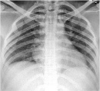
Table 13.
Clinical input by radiologist for misclassified images.
| Images | Ground truth | Prediction | Clinical input |
|---|---|---|---|
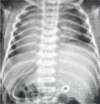 |
COVID-19 | Normal | X-ray image of the pediatric patient has less filed of the lung than mediastinum, so the software learning algorithm picks up as normal (healthy). |
 |
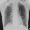
COVID-19 | Normal | No explanation has to correlate with chest auscultation findings. |
 |
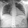
Normal | COVID-19 | X-ray image has an area of retro cardiac opacity and cardiac silhouettes deviation, so the software learning algorithm may have picked up as COVID-19. |
 |
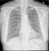
Bacterial Pneumonia | COVID-19 | X-ray image has hilar lymph nodes and peripheral opacity, so the software learning algorithm may have picked up as COVID-19. |
 |
COVID-19 | Normal | No explanation has to correlate with chest auscultation findings |