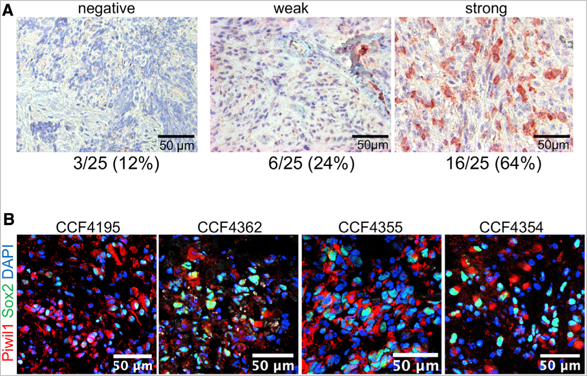
Figure 1. Piwil1 Is Frequently Overexpressed in GBM.

(A) Immunohistochemical staining of Piwil1 in human GBM.
(B) Immunofluorescence staining of Piwil1 (red) and Sox2 (green) in human GBM. Nuclei were counterstained with DAPI (blue).
See also Figure S1.
