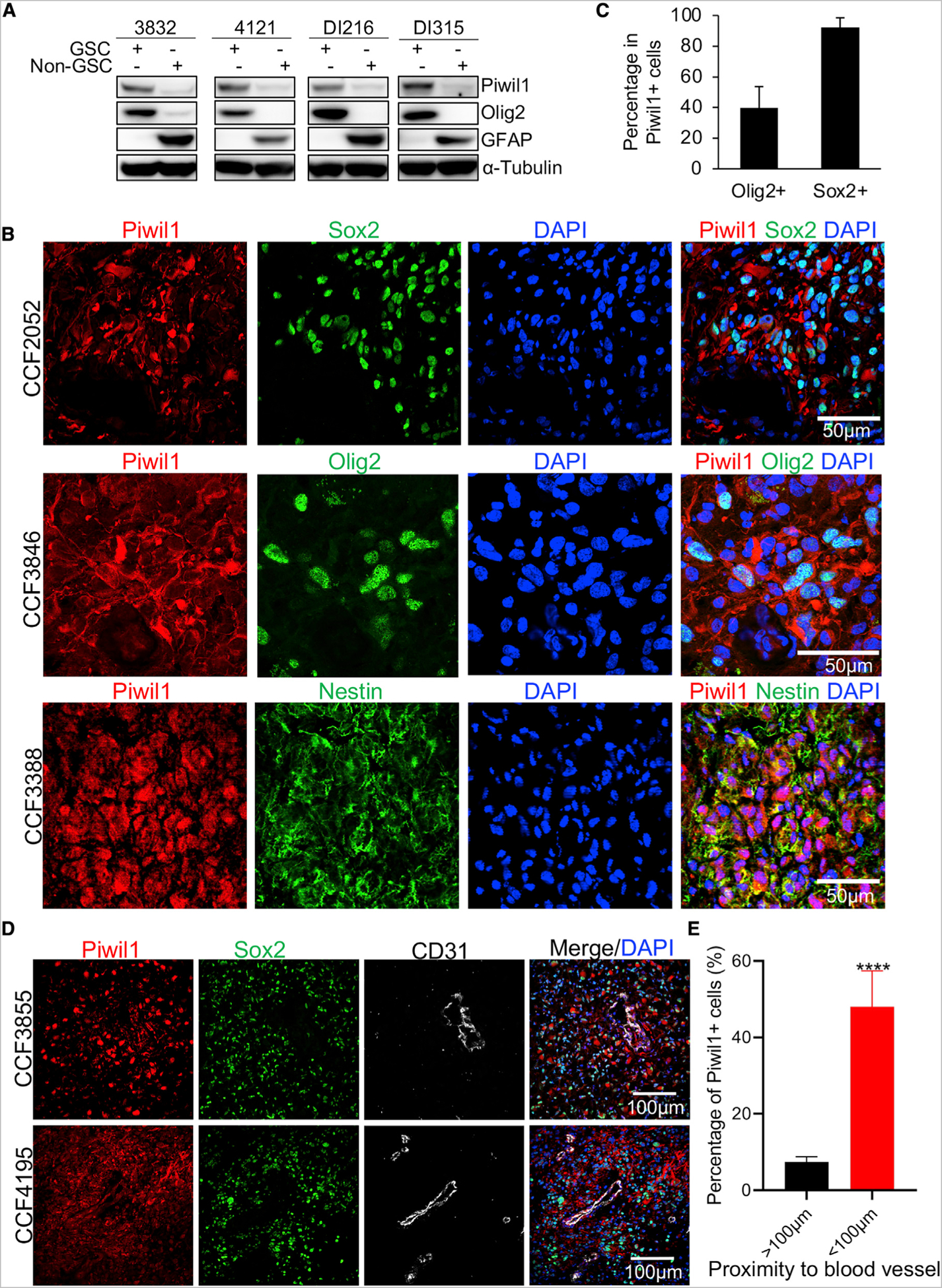
Figure 2. Piwil1 Is Preferentially Expressed in GSCs.

(A) Western blots of Piwil1, stem cell maker Olig2, and astrocyte marker GFAP in four paired GSCs and non-GSCs.
(B) Immunofluorescence imaging of Piwil1 (red) and stem cell markers (green) Sox2 (top), Olig2 (middle), and Nestin (bottom) in human GBM specimens. Nuclei were counterstained with DAPI (blue).
(C) Quantification of percentages of Sox2+ (n = 9 high-powered fields) and Olig2+ (n = 9 high-powered fields) cells in Piwil1+ cells.
(D) Immunofluorescence imaging of Piwil1 (red), Sox2 (green), and endothelial cell marker CD31 (gray) in human GBM specimens. Nuclei were counterstained with DAPI (blue).
(E) Quantification of Piwil1+ cells percentage within and beyond 100 μm distance from blood vessels (n = 15 high-powered fields).
****p < 0.0001. Data are represented as mean ± SD. See also Figure S2.
