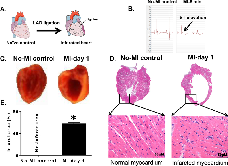
Figure 1. MI-induced ST-elevation and structural heart failure pathology in mice.
(A).Schematic figure indicating coronary artery ligation point for surgery to induce myocardial infarction (MI). (B) Ligation-induced ST-elevation indicated successful MI surgery compared to no-MI controls. (C). Digital photograph of the vertical half-cut left ventricle (LV) stained with 2,3,5- triphenyltetrazolium chloride (TTC) following permanent coronary ligation indicating the infarcted area (white) and the non-infarcted remote area (red) compared to no-MI naïve controls. (D). Mice LV stained with hematoxylin (H) and eosin (E) post-MI. LV (1.25x) and amplified photographs (40x) of no-MI naive control indicate healthy myocardium and post-MI histology indicate signs of myocardial damage from the ligation-induced injury. (E). Graph is indicating the quantitative measurement of ligation-induced infarct area after coronary ligation versus non-ligated no-MI controls. *p<0.05 vs no-MI naïve controls (n=6 mice/group).

