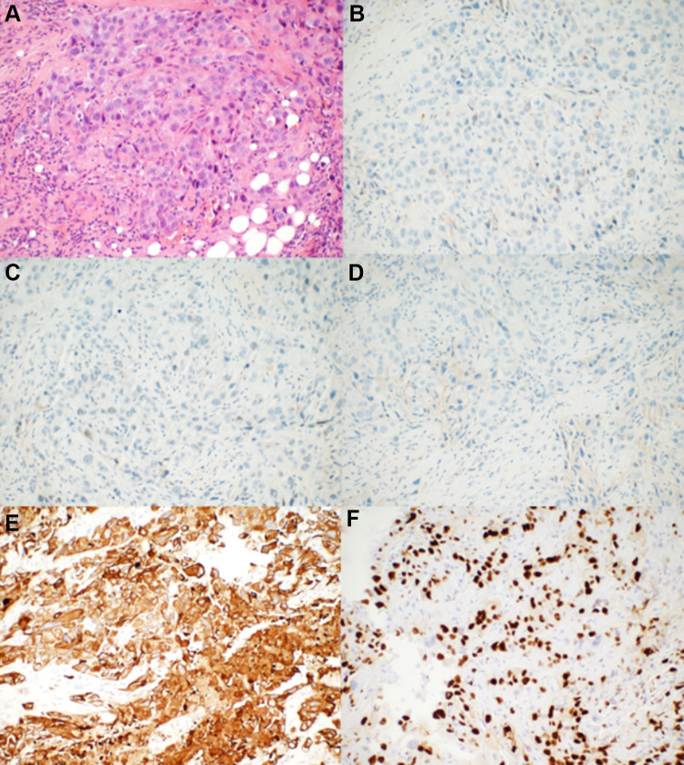
Figure 1.
The pathological results of the rebiopsy pathology of the right breast tumor performed in our hospital. (A) Hematoxylin–eosin-stained sections revealed that the tumor cells grew in a solid and patchy infiltrating manner (original magnification: 200×). (B–D) ER, PR, and HER-2 were negative for neoplastic cells by immunohistochemical analysis (original magnification: 200×). (E) CK5/6 was strongly expressed by tumor cells (original magnification: 200×). (F) Ki-67 was expressed in the nuclei of approximately 50% of tumor cells (original magnification: 200×).

