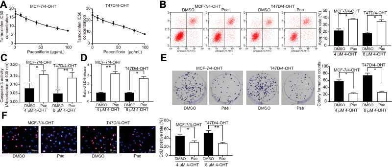
Figure 2.
Pae inhibits 4-OHT resistance in BC cells. (A) The IC50 values of 4-OHT on MCF-7/4-OHT or T47D/4-OHT cells under Pae exposure (0–100 μg/mL) examined by CCK-8 assays. Pae at 100 μg/mL was used for the later experiments. (B) The apoptosis levels of MCF-7/4-OHT and T47D/4-OHT cells under Pae exposure (100 μg/mL) detected by flow cytometry under treatment of 4 μM and 8 μM 4-OHT, respectively; (C) the apoptosis levels of MCF-7/4-OHT and T47D/4-OHT cells under Pae exposure (100 μg/mL) assessed by Caspase-3 activity kit; (D) the cytotoxicity of 4-OHT to MCF-7/4-OHT and T47D/4-OHT cells under Pae exposure (100 μg/mL) determined by a LDH kit; (E) colony formation of MCF-7/4-OHT and T47D/4-OHT cells under Pae exposure (100 μg/mL) tested by colony formation assays; (F) number of S-phase cells under Pae exposure (100 μg/mL) detected by EdU assays. Data are displayed in the form of mean ± SD. All experiments were repeated at least three times. In panel (A) two-way ANOVA along with Tukey’s multiple comparison was applied, while in panel (B–F) one-way ANOVA along with Tukey’s multiple comparison was used. *p < 0.05, **p < 0.01.
Abbreviations: Pae, paeoniflorin; BC, breast cancer; IC50, median inhibition concentration; 4-OHT, 4-hydroxytamoxifen; CCK-8, cell counting kit-8; LDH, lactose dehydrogenase; EdU, 5-ethynyl-2ʹ-deoxyuridine; SD, standard deviation; ANOVA, analysis of variance.

