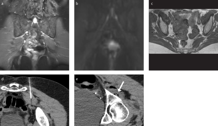
Figure 5. a–e.
A 78-year-old female presenting with buttock pain and back pain and diagnosed with left piriformis syndrome. She received an injection without Botox and had a negative response. Coronal 3D IR TSE image (a) shows normal signal of the sciatic nerves. Coronal MIP DTI image (b=600 s/mm2) (b) shows no enhancement of the sciatic nerves. Axial T1-weighted image (c) shows left piriformis hypertrophy. CT-guided injection (d) without Botox of the left piriformis muscle. Post-injection image (e) of the sciatic nerve (black arrow) and medication mixture (white arrow).

