
Figure 7.
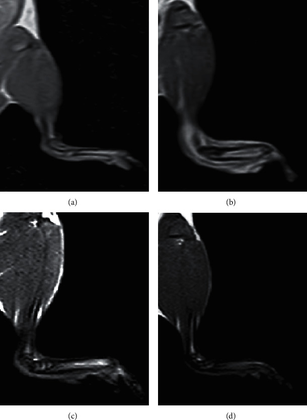
T1-weighted MR images of the right ankle joints of normal control, MIA, and MIA + PRP groups showing normal joint and foot anatomy (Figure 7(a)), enlarged diameter of the joint with extensive soft-tissue edema in acute osteoarthritic rats (Figure 7(b)), and reduced soft tissue edema in chronic osteoarthritic rats and still enlarged joint diameter as compared to the normal control (Figure 7(c)). In contrast, PRP treatment revealed a diminished diameter of the joint resembling that of normal control (Figure 7(d)). (a) Normal control. (b) Acute OA (MIA group). (c) Chronic OA (MIA group). (d) MIA-PRP group.