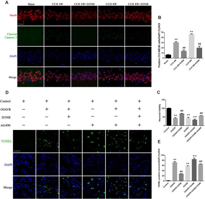
Figure 6.
D3NB protects hippocampal neurons by antagonizing the action of apoptosis in the CCH rats and OGD/R environment in vitro. (A) Double staining of NeuN and cleaved Caspase 3 in apoptotic neurons in the CA1 region of the hippocampus in different groups. (B) The positive cell numbers were quantified by ImageJ Software (magnification ×60, scale bar = 30 μm. **P < 0.01 CCH groups vs. Sham; ##P < 0.01 CCH + D3NB groups vs. CCH groups; n = 3/group, quantitative analysis of ROI, 200 μm2). (C) Cell viability was measured using the CCK-8 method following treatment with AG490 and/or D3NB after exposure to OGD/R in vitro (OGD/R, OGD 2 h/R 48 h. **P < 0.01 compared to Control; **’P < 0.01 OGD/R + AG490 vs. AG490; ##P < 0.01 OGD/R+D3NB vs. OGD/R; ##’P < 0.01 OGD/R + D3NB + AG490 vs. OGD/R+AG490. n = 5/group). (D) TUNEL and DAPI staining of primary hippocampal neurons in the control, OGD/R, OGD/R+D3NB, control+AG490, OGD/R + AG490, and OGD/R + AG490 + D3NB groups. (E) Quantitative analysis (OGD/R, OGD 2 h/R 48 h. Magnification ×60, scale bar = 30 μm. **P < 0.01 compared to Control; **’P < 0.01 OGD/R + AG490 vs. AG490; ##P < 0.01 OGD/R + D3NB vs. OGD/R; ##’P < 0.01 OGD/R + D3NB + AG490 vs. OGD/R + AG490. n = 3/group, quantitative analysis of ROI, 200 μm2).

