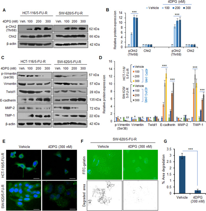
Fig. 2. 4DPG activates Chk2 and attenuates EMT in 5-FU-R CRC cells.
A Western blotting analysis results portraying protein expression of p-Chk2 (Thr68) and Chk2 in HCT-116/5-FU-R and SW-620/5-FU-R cells in response to increasing concentrations of 4DPG, treated for 48 h. β-Actin expression was considered as endogenous loading control. B Densitometry analysis showing relative protein expression of western blot bands presented above. n = 3, error bars: mean ± SD; ***P < 0.001. C Western blots depicting protein expression of EMT markers in HCT-116/5-FU-R and SW-620/5-FU-R cells in response to increasing concentrations of 4DPG, treated for 48 h. β-Actin expression was considered as endogenous loading control. D Densitometry analysis of western blots presented in (C) showing quantification of protein expression. n = 2, error bars: mean ± SD; *P < 0.05, **P < 0.01, ***P < 0.001. E Immunocytochemistry results displaying the protein expression levels of Vimentin in HCT-116/5-FU-R and SW-620/5-FU-R cells, treated with vehicle and/or 4DPG (300 nM) for 48 h. Scale bars: 50 µm. F In situ fluorescent gelatin degradation assay results showing the invadopodia/footprints of invaded SW-620/5-FU-R cells treated with vehicle and/or 4DPG at the indicated concentration for 48 h. Blue stains over the green background display DAPI staining of nuclei of cells. G Quantification of the area of degradation through Image J software. n = 3, error bars, mean ± SD; ***P < 0.001.

