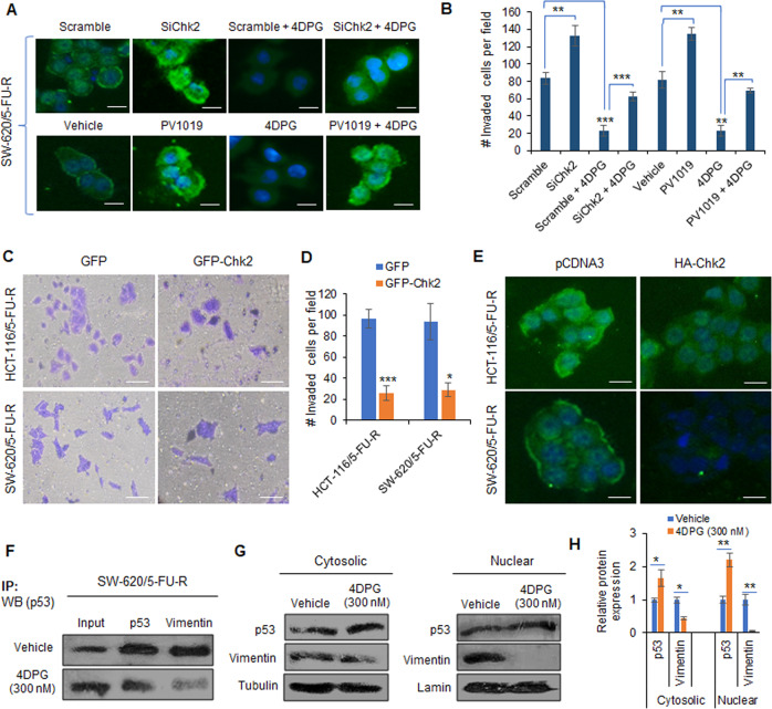
Fig. 3. Chk2-dependent role of 4DPG in modulating p53 and repressing Vimentin expression in 5-FU-R cells.
A Immunocytochemistry results showing the Vimentin expression level in SW-620/5-FU-R cells treated/transfected with scramble, SiChk2, scramble plus 4DPG, SiChk2 plus 4DPG, vehicle, PV1019, 4DPG, and PV1019 plus 4DPG for 48 h. Scale bars: 50 µm. B Quantification of number of invaded HCT-116/5-FU-R cells exposed to the above treatment conditions for 48 h. n = 2, error bars, mean ± SD; **P < 0.01, ***P < 0.001. C Matrigel invasion assay results showing invaded HCT-116/5-FU-R and SW-620/5-FU-R cells following 48 h of transient transfection with GFP and GFP-Chk2 plasmid construct. Scale bars: 50 µm. D Quantification of the number of invaded cells per field from the above experimental conditions. n = 3, error bars, mean ± SD; *P < 0.05, ***P < 0.001. E Immunocytochemistry results displaying Vimentin expression levels in HCT-116/5-FU-R and SW-620/5-FU-R cells after 48 h of transient transfection with pCDNA3 and/or HA-Chk2 plasmid construct. Scale bars: 50 µm. F Immunoprecipitation with Vimentin or p53 followed by western blotting analysis of p53 in SW-620/5-FU-R cells treated with vehicle and/or 4DPG (300 nM) for 48 h. G Cytosolic (left panel) and nuclear (right panel) extracts of SW-620/5-FU-R cells treated with vehicle and/or 4DPG (300 nM) for 48 h showing protein expression of Vimentin and p53. Tubulin and lamin expression was considered as endogenous loading controls for cytosolic and nuclear fractions, respectively. H Densitometry analysis of the above western blots showing relative protein expression of p53 and Vimentin in cytoplasmic and nuclear extracts. n = 3, error bars: mean ± SD; *P < 0.05, **P < 0.01.

