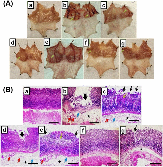
Figure 9.
(A) Macroscopically investigation of mucosal lesions in glandular part of the stomachs. (a) N group, (b) ulcer group, (c) ome group, (d) plain group, (e) ALL group, (f) NanoAL group, and (g) NanoAH group. (B) Microscopical examination of glandular part of stomach sections stained by (H&E) (X: 100). (a) N group, (b) ulcer group, (c) ome group, (d) plain group, (e) ALL group, (f) NanoAL group, and (g) NanoAH group. Green arrow points to visual changes than the normal mucosa present in N group, thick black arrow points to mucosal ulceration, thin black arrow points to erosion, yellow arrow points to necrosis, asterisk points to edema, red arrow points to dilated blood vessel, and blue arrow points to leukocytic cells infiltration.

