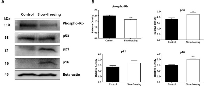
Figure 5.
Western blot analysis of phospho-pRb, p53, p21, and p16INK4a in slow-frozen and control ovarian tissues. (A) Based on the senescence protein marker p16, four related proteins were measured in the same human ovarian tissue. Parallel blots were probed with β-actin antibodies as a protein quantification control. Full-length blots are presented in Supplementary Figure 1. (B) Graphs of the fold-change value from the control after quantification of the western blot analysis results. Images were acquired with the ChemiDoc Imaging Systems (BIO-RAD) and band intensities were quantified with ImageJ software. Paired Student's t-test was used in these analyses. *p-value < 0.05. **p-value < 0.005.

