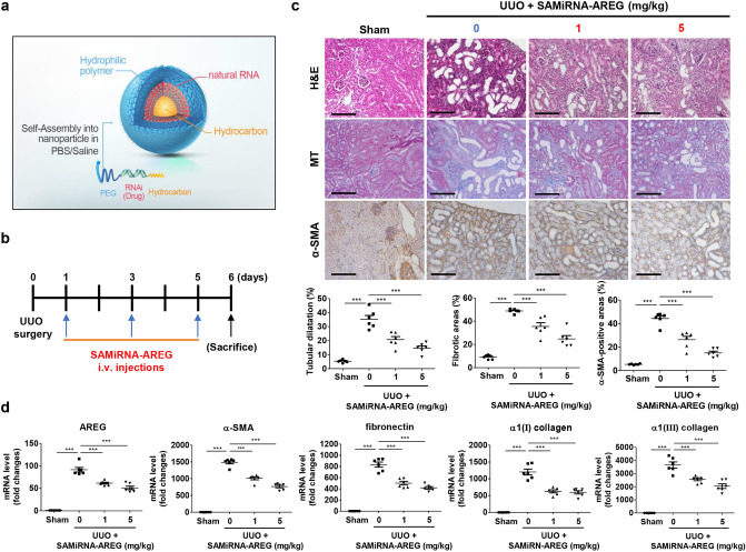
Figure 3.
SAMiRNA-AREG ameliorated kidney fibrosis in the UUO-induced model. (a) The structure of SAMiRNA-AREG. (b) Time scheme for SAMiRNA-AREG efficacy analysis in UUO mice. (c) Histological analysis by H&E, MT, and IHC staining of control, UUO, and SAMiRNA-AREG-treated UUO mice. Tubular dilatation, the fibrotic areas, and the α-SMA-positive areas were quantified. Scale bar, 100 μm. (d) Kidney tissue lysates were subjected to real-time qRT-PCR analysis for AREG, α-SMA, fibronectin, α1(I) collagen, and α1(III) collagen. ***p < 0.001 compared with UUO mice by ANOVA with the Newman-Keuls post-hoc test.

