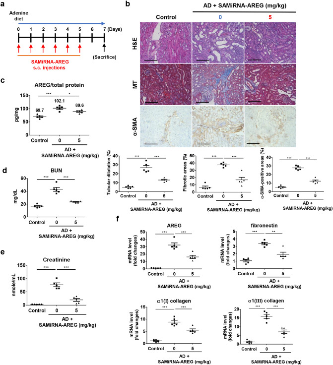
Figure 4.
SAMiRNA-AREG ameliorated renal fibrosis in the AD-induced model. (a) Time scheme for SAMiRNA-AREG efficacy analysis in AD mice. (b) Histological analysis by H&E, MT, and IHC staining of control, UUO, or SAMiRNA-AREG-treated AD mice. Tubular dilatation, fibrosis areas, and α-SMA-positive areas were quantified. Scale bar, 100 μm. (c) AREG proteins in kidney tissue lysates were measured by ELISA. The numbers indicate the mean value of each data set. (d,e) BUN and serum creatinine were measured by ELISA. (f) Kidney tissue lysates were subjected to real-time qRT-PCR analysis for AREG, fibronectin, α1(I) collagen, and α1(III) collagen (mean ± SEM). *p < 0.05, **p < 0.01, ***p < 0.001 compared with AD mice by ANOVA with the Newman-Keuls post-hoc test.

