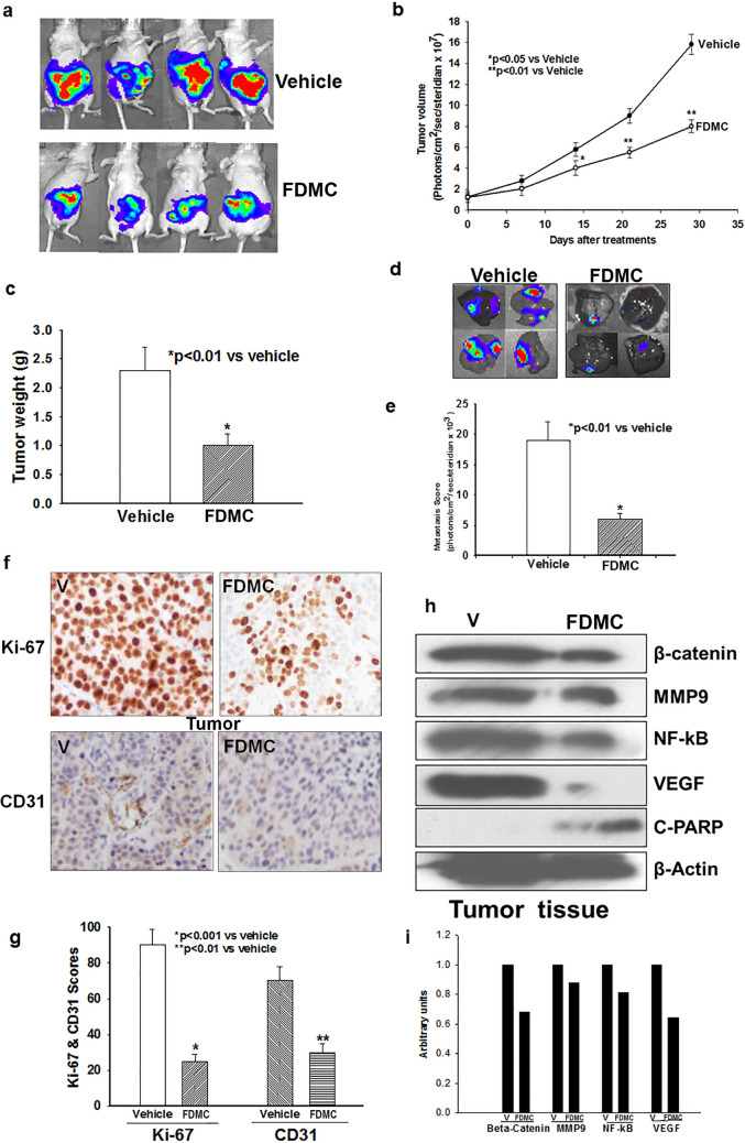
Figure 6.
Effect of FDMC on colorectal tumour growth, metastasis, inflammation, and angiogenesis markers, β-catenin expression and induction of apoptosis in mice, as evidenced by comparisons with vehicle-treated controls (V = vehicle). (a) Luciferase bioluminescence image shows that treatment with FDMC (200 mg/kg, orally, twice a week) for 4 weeks decreased tumour growth in mice. (b) FDMC significantly decreased tumour volume 2 weeks after treatment (*p < 0.05) and more significantly (**p < 0.01) after 3 to 4 weeks of treatment. (c) FDMC significantly decreased tumour weight 4 weeks after treatment (*p < 0.01). (d) Luciferase bioluminescence image shows that FDMC decreased the numbers of liver metastasis nodules in mice. (e) FDMC significantly decreased liver metastases (luminescence units) mice (*p < 0.01). (f,g) Histology data of colorectal tumours show that FDMC significantly inhibited tumour cell proliferation (Ki-67) and tumour angiogenesis (CD31). (h) Western blot data depict the depletion of markers for inflammatory, angiogenesis, and metastasis (NF-kB, VEGF and MMP9) and induced apoptosis (C-PARP) in the tumour tissue of mice treated with FDMC compared with vehicle. Results are mean ± SEM (bars; n = 3–5).

