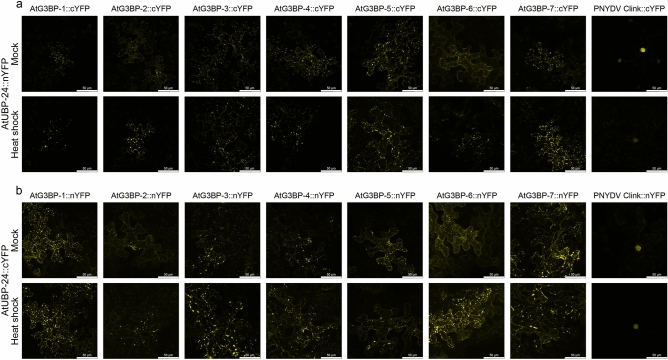
Figure 4.
Visualization of protein–protein interaction of the AtUBP-24 with the various AtG3BPs and PNYDV Clink via bimolecular fluorescence complementation (BiFC) under ambient conditions and heat shock (45 min at 37 °C). (a) Interaction of AtUBP-24 with the corresponding partners fused C-terminally with splitYFP. (b) Interaction of AtUBP-24 and the respective AtG3BPs and PNYDV Clink N-terminally fused to splitYFP. All pictures are projections of z-stacks obtained by confocal laser scanning microscopy. The individual pictures correspond to a size of 185 × 185 µm.

