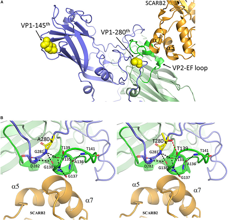
FIGURE 3.
The mutant threonine of VP1-280T is located at the interaction hub between VP1, VP2 and the entry receptor hSCARB2. (A) VP1 amino acid 280, but not amino acid 145, is near the contact site between EV-A71 and hSCARB2, according to the cryoEM structure of the binding complex of VP1, VP2 and hSCARB2 at low pH (Zhou et al., 2019). PDB accession code: 6I2K. Orange, human SCARB2; Blue, VP1 capsid protein; Green, VP2-EF loop; Yellow, VP1 amino acids 145 and 280. (B) A loop turn of the VP2-EF loop (green) is in contact with the α7 helix of SCARB2 (orange). The backbone atoms of VP1 and the VP2 EF loop are represented as sticks with carbon atoms colored in light blue and green, respectively. This VP2 loop turn can be stabilized by a hydrogen-bonded network (black dashed lines) between the backbone oxygen and nitrogen atoms of VP1 G281-D282 and those of VP2 V135-T141. One de novo created hydrogen bond between the oxygen atom (hydroxyl group) of the mutant VP1-280T and the carbonyl group of VP2-139T is colored in a red dashed line. Residues A280 and T280 of VP1 are shown in sticks with carbon atoms in yellow. This figure is produced using Discovery Studio Visualizer and PyMOL (see section “Materials and Methods”).

