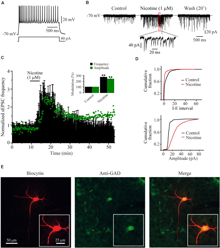
FIGURE 1.
Nicotine increased the frequency and evoked bursts of sEPSCs recorded from RMTg GABAergic neurons. (A) Voltage response of a typical GABAergic RMTg neuron to a depolarizing current step of 40 pA. (B) Current traces showing sEPSCs recorded in voltage clamp mode from the same cell in A. Control condition (left), 10 min after nicotine (middle) and 20 min after nicotine washout (right). A region of the middle trace (red box) is displayed below at a slower sweep time where nicotine-evoked bursts of sEPSCs are evident. (C) Time-frequency histogram (n = 15) shows the temporal course of nicotine effect. The graph in green shows the time course of the sEPSC amplitude. For clarity, the errors were removed from the graph. The summary of the 15 cells is shown in the bar graph (inset) (Mann–Whitney U-test). (D) Cumulative fraction plots (cell in A,B) of the inter-event interval (I–E, top) and amplitude (bottom) showing that nicotine increased both the frequency and the amplitude of sEPSCs. (E) Same cell in A and B labeled with biocytin (left), anti-GAD65/67 antibody (middle), and merge (right). All the experiments were performed in the presence of bicuculline (10 μM). **p < 0.01.

