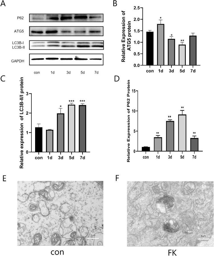
Figure 3.
Autophagy flux was impaired in Fusarium solani keratitis. (A–D) The ratio of LC3II/I and P62 protein expression levels were increased significantly in the F. solani-infected mouse corneal tissues and reached a peak on day 5, whereas the ATG5 protein expression levels were decreased at the fifth day. (E, F) Transmission electron microscopy (TEM) showed autophagosomes in both groups at day 5. n = 6; *P < 0.05, **P < 0.01, ***P < 0.001.

