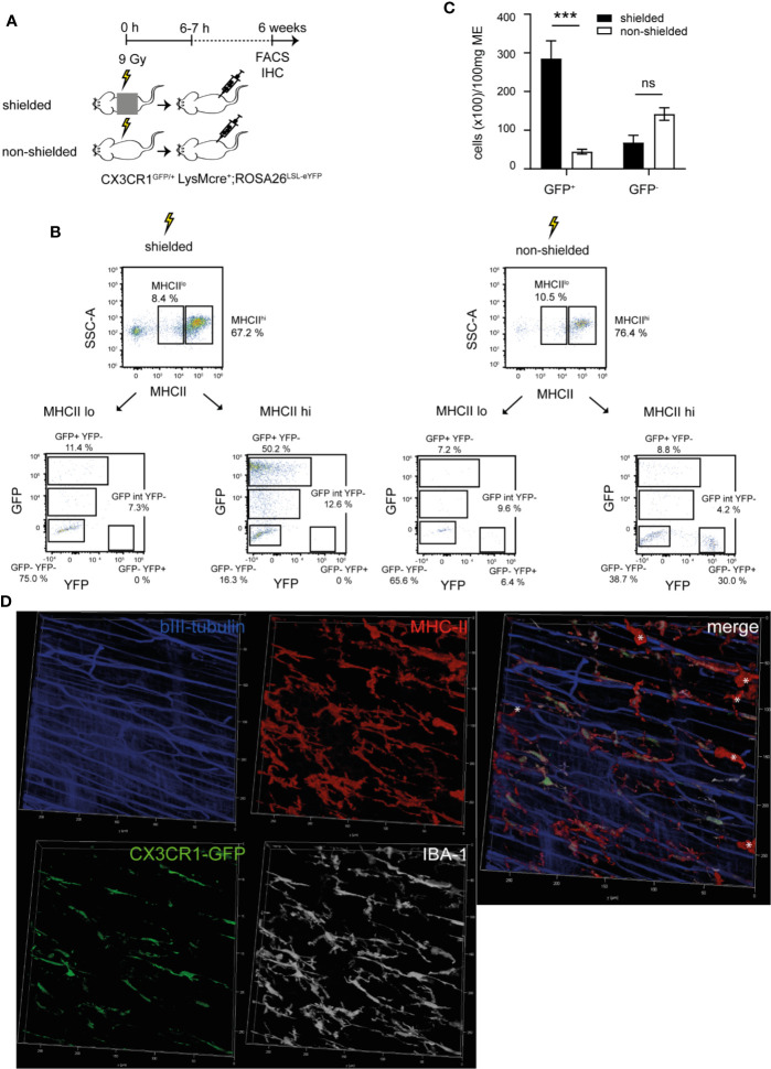
Figure 5.
Radio-resistant MHCIIhiCX3CR1- cells express the macrophage marker IBA-1 and reside within a special anatomical niche. (A–C) Lethally irradiated shielded and non-shielded CX3CR1GFP/+ were recovered 6–7 h after radiation with a total of 1.2 x 10⁷ bone marrow (BM) cells of LysMcre+;ROSA26LSL-eYFP donor mice. Six weeks later, fluorescence activated cell sorting (FACS) analysis and whole mount immunohistochemistry (IHC) was performed. (A) Scheme of the experimental setup. (B) Representative FACS plots of the expression of GFP (CX3CR1)/YFP (LysM) on living CD45+ MHCIIhi/MHCIIlo gated cells isolated from the ME with Invitrogen™ Attune™ NxT in shielded and non-shielded CX3CR1GFP/+ x LysMcre+;ROSA26LSL-eYFP and (C) quantification of total CD45+MHCIIhiGFP+ and CD45+MHCIIhiGFP- cells. (D) Representative confocal image stack of CX3CR1GFP/+ stained for GFP (green), MHCII (red), βIII-tubulin (blue) and IBA-1 (gray) revealing the existence of a MHCIIhiIBA-1+CX3CR1- cell population (*) located in a different layer than the MHCIIhiIBA-1+CX3CR1+ cells. Experiments were performed three times reaching an overall size of n = 3 animals per group. Means + SEM were generated from the overall numbers of each group. Samples were analyzed by student’s t-test and the results are displayed as means ± SEM. ***p ≤ 0.001. For images on the localization, please refer to Supplementary Figure S7 .

