Figure 7.
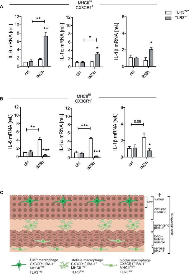
Toll-like receptor 3 (TLR3) signaling in MHCIIhiCX3CR1- cells drives cytokine expression during POI. Wt and TLR3-/- mice underwent IM or were left untreated (ctrl). Three h after surgery (IM3h), gene expression levels of IL-6, IL-1β, and IL-1α were analyzed on (A) MHCIIhiCX3CR1- and (B) MHCIIhiCX3CR1+ cell populations that had been sorted beforehand from the ME samples via fluorescence activated cell sorting (FACS) and displayed as fold changes after normalization to ctrl. n = 3 (each replicate was pooled from six mice). Samples were analyzed by two-way analysis of variance with Tukey post hoc test and student’s t-test, respectively, and the results are displayed as means ± SEM. Experiments were performed three times reaching an overall size of n = 3 animals per group. Means + SEM were generated from the overall numbers of each group. *p ≤ 0.05, **p ≤ 0.01, ***p ≤ 0.001 versus the corresponding TLR3+/+ group or indicated groups. (C) Schematic drawing of the intestinal muscularis externa (ME) illustrating the location of bipolar or stellate MHCIIhiCX3CR1+ macrophages and MHCIIhiCX3CR1- macrophages. The latter reside within a distinct anatomical niche, the deep myenteric plexus (DMP), and express higher TLR3 levels while the MHCIIhiCX3CR1+ cells are located within the myenteric and serosal plexus and show low TLR3 expression. Intestinal manipulation (IM) triggers a proinflammatory cytokine expression during the onset phase of postoperative ileus (POI) mainly in MHCIIhiCX3CR1- macrophages and this mechanism depends on TLR3 signaling.

