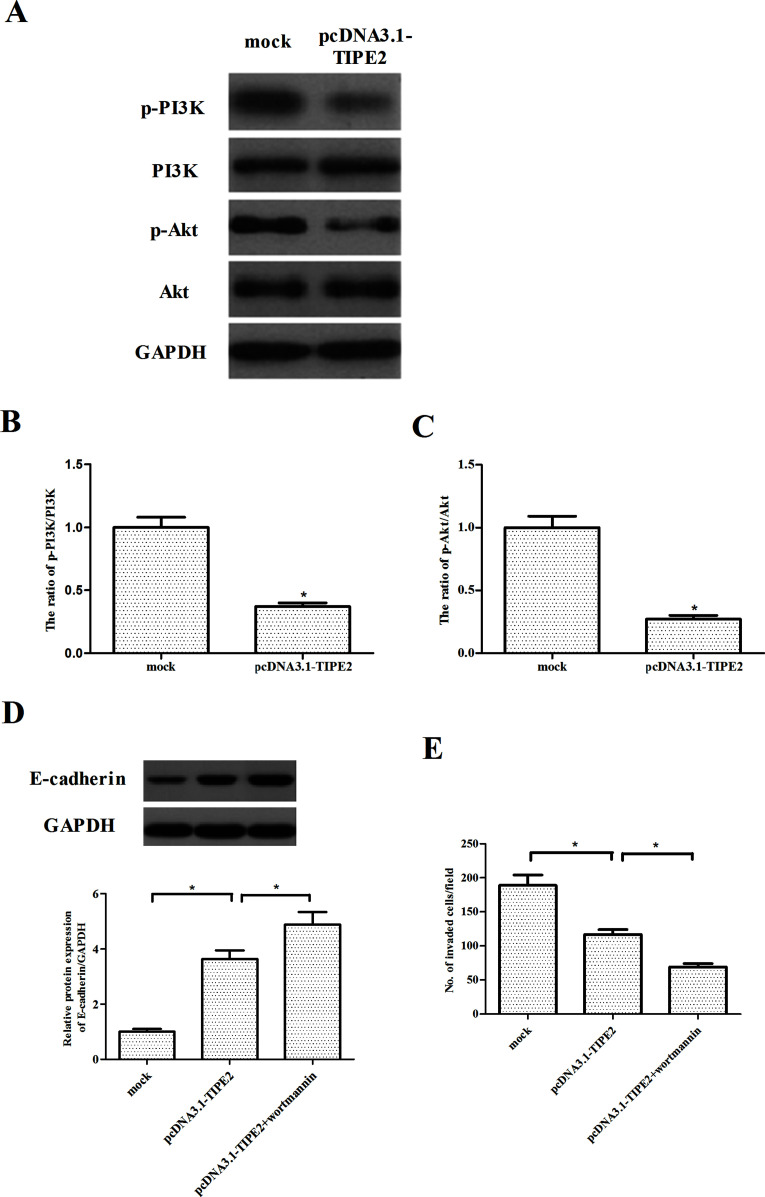
Figure 5.
TIPE2 inhibits the PI3K/Akt signaling pathway in prostate cancer cells. PC-3 cells were transfected with pcDNA3.1-TIPE2 or mock for 24 h. (A) The expression of p-PI3K, PI3K, p-Akt, and Akt proteins was detected by Western blotting. GAPDH served as a loading control. (B, C) Quantification analysis was performed using Gel-Pro Analyzer version 4.0 software. (D) PC-3 cells were transfected with pcDNA3.1-TIPE2 or mock in the presence or absence of wortmannin (100 nM) for 24 h. The expression of E-cadherin was analyzed via Western blotting. (E) Cell invasion was evaluated by the Transwell invasion chamber assay. Data are mean ± SD values from three experiments, each performed in triplicate. Compared with the mock group, *p < 0.05.

