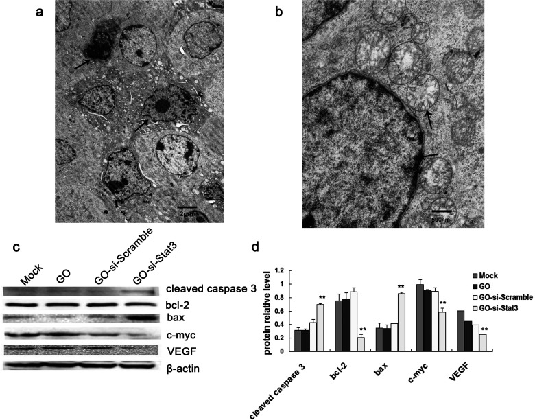
Figure 5.
(a) Transmission electron microscope image of B16 cells treated with GO-si-Stat3. The arrows indicate typical apoptotic cells. (b) TEM image of B16 cells treated with GO-si-Stat3. The upper arrow indicates fractured mitochondrial cristaes. The lower arrow indicates chromatin margination. (c) Western blot method of investigation of Stat3-related proteins in B16 cells. (d) Calculation of relative Stat3-related protein expression in each group. **p < 0.01 versus mock, GO, and GO-si-Scramble.

