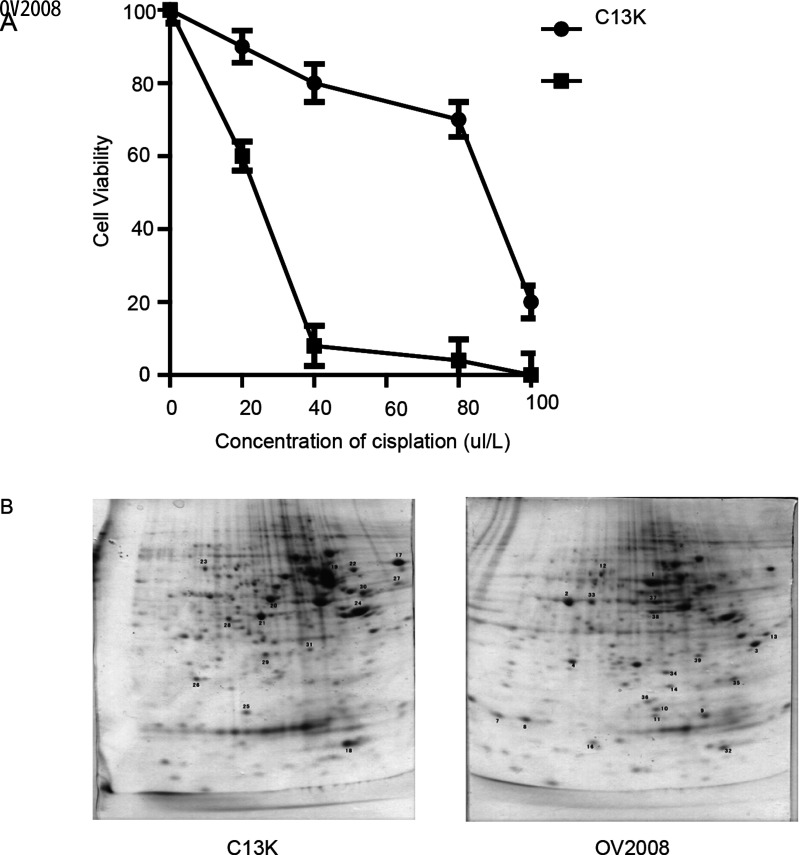
Figure 1.
Cisplatin-induced cytotoxicity and apoptosis in resistant (C13*) and sensitive cell lines (OV2008) and 2-DE gel electrophoresis results. (A) Cells were cultured in the presence of the indicated concentrations of cisplatin for 48 h. Cell viability in response to treatment with escalating doses of cisplatin was assessed by CCK-8. (B) Representative image of 2-DE urea gel electrophoresis of human ovarian cancer cells. Proteins were detected by Coomassie blue staining on the 2-DE images of the master gel.

